Diagnosing And Treating A Cholesteatoma
Magnetic Resonance Imaging
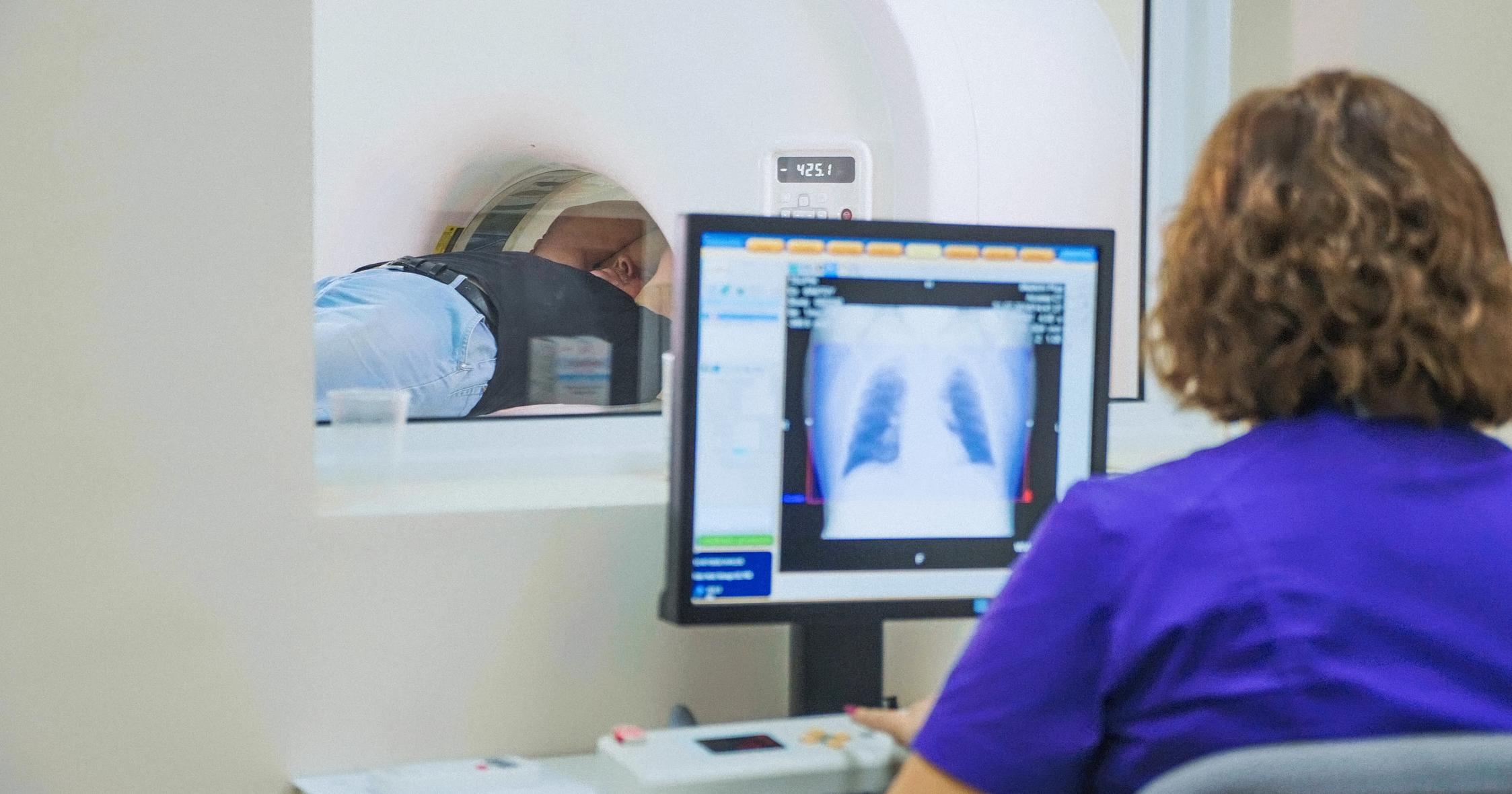
If a CT scan determines further imaging is required, magnetic resonance imaging can provide even more detail. Unlike a CT scan, an MRI does not use X-rays instead using radio waves and a magnetic field. This technique is noninvasive and can be especially useful for pre- and post-operation. A person is placed within the MRI scanner, and a 3-D image is produced without the use of harmful radiation. This can show precisely where the excess skin of the cholesteatoma is growing, how deep it has progressed, and help to determine the best way to remove it. Using magnetic resonance imaging post-operation allows doctors to determine if the area is healing properly, has developed an infection, or if scar tissue has formed.
Learn more about treating a cholesteatoma now.
Mastoidectomy

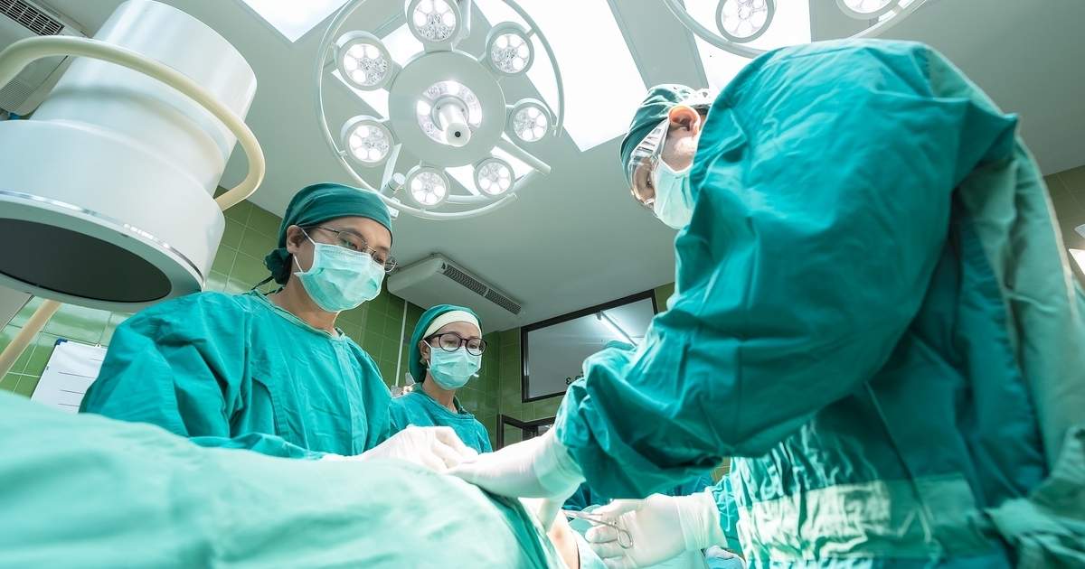
A surgeon performs a mastoidectomy to remove the air cells of the mastoid process, the bone directly behind the ear. This is often the best way to remove a cholesteatoma that continually causes infection. The incision is made in the valley behind the ear, and the cholesteatoma can be removed. Depending on how much it has grown and how much damage has been caused to the inner ear, the bones of the inner ear can be repaired or reconstructed. They can also be replaced with prosthetic bones. The main goal of the surgery is to remove any infection, but often hearing is improved after the cholesteatoma has been fully removed. Complications can include pain and infection, though this is rare. The ability to taste can be altered if the nerve near the ear is affected during surgery.
This is often performed before a tympanoplasty, which is coming up next.
